Three medical-technology studies published in March 2026 reported useful results in colorectal surgery, stroke care and advanced heart failure. They did not test one shared artificial-intelligence system, and they did not establish that AI broadly reduces surgical mortality, restores mobility after stroke or diagnoses heart failure before symptoms.
The colorectal evidence concerned indocyanine green fluorescence angiography, an imaging technique that helps surgeons assess blood flow before joining sections of bowel. The stroke trial evaluated a package combining AI-assisted scan analysis, classification of stroke causes and treatment recommendations. The heart-failure study trained a model to predict a measurement normally obtained during an exercise test.
Reading the studies together is possible only if their endpoints remain separate. One analyzed an operative complication, one measured recurrent vascular events and care processes, and one evaluated prediction accuracy. Each result answers a different clinical question.
The Colorectal Meta-analysis Tested Fluorescence Imaging, Not AI
An anastomotic leak can occur when a surgical join in the bowel fails to heal. Indocyanine green fluorescence angiography, or ICGFA, gives surgeons a near-infrared view of tissue perfusion during an operation. That information can prompt a surgeon to change where the bowel is divided or joined.
The Lancet Gastroenterology & Hepatology review pooled nine randomized controlled trials involving 4,754 patients. The authors reported that ICGFA was associated with about a 40% relative reduction in anastomotic leakage, with the clearest evidence in left-sided and rectal colorectal surgery.
That is a clinically important result for the complication actually measured. It does not show that the technique reduced mortality, infection, readmission or recovery time unless those outcomes were separately demonstrated. A relative reduction also does not tell a patient the absolute change in risk without the baseline event rate for the relevant operation and population.
ICGFA is a digital visualization and decision-support technique, but the meta-analysis did not test an AI algorithm interpreting the images. The researchers said the established imaging principle could support future AI tools. Turning that future direction into a claim that an AI system already produced the trial benefit assigns the evidence to the wrong technology.
The Stroke Trial Reduced a Composite Vascular Endpoint
The GOLDEN BRIDGE II trial enrolled 21,603 adults with acute ischemic stroke across 77 hospitals in China. Hospitals, rather than individual patients, were randomized. Thirty-eight hospitals used the clinical decision-support system for 11,054 patients, while 39 hospitals provided usual care to 10,549 patients.
The intervention was broader than fast scan reading. It combined AI-assisted magnetic-resonance-image analysis, classification of stroke cause and evidence-based treatment recommendations integrated into hospital systems. Staff at intervention hospitals also received training. The trial therefore tested the performance of an implemented care package, not an isolated algorithm operating independently of clinicians.
At three months, a new vascular event occurred in 2.9% of the intervention group and 3.9% of the control group. The composite included ischemic or hemorrhagic stroke, myocardial infarction and vascular death. At 12 months, the rates were 4.0% and 5.5%, respectively. Adherence to a composite set of evidence-based care measures was also slightly higher with decision support, at 91.4% versus 89.8%.
The trial did not find significant differences in disability or all-cause mortality at three, six or 12 months. Moderate or severe bleeding and overall bleeding also did not differ significantly. It is therefore accurate to report fewer composite vascular events under the intervention, but inaccurate to convert that result into proven recovery of mobility, lower mortality or a general reduction in disability-related costs.
Because randomization occurred by hospital, local practice patterns and post-discharge care could have influenced the result. The large, pragmatic design is a strength, while the combined intervention makes it difficult to attribute the entire effect to scan analysis alone. Testing the system in health systems outside China would address how well the result travels across workflows and patient populations.
The Heart Model Predicted an Exercise-Test Measure
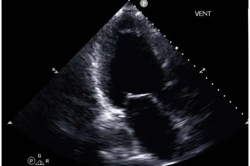
Advanced heart-failure assessment can use cardiopulmonary exercise testing, or CPET, to measure peak oxygen consumption. CPET requires specialized equipment and staff. The npj Digital Medicine study asked whether a model could estimate peak oxygen consumption from more readily available echocardiography images and structured electronic-health-record data.
Researchers developed the model with records from 1,000 patients at one NewYork-Presbyterian site and evaluated it externally in 127 patients from three other affiliated campuses. On the development test set, the model achieved an R-squared value of 0.603 for peak oxygen-consumption prediction and an area under the receiver-operating-characteristic curve of 0.849 for identifying patients below a high-risk threshold. On the external cohort, the corresponding results were 0.541 and 0.870.
Those are model-performance measures, not proof of diagnosis or benefit to patients. The retrospective study included people who had been referred for CPET, all four hospitals were academic centers in the New York metropolitan area, and the external cohort was small. The ultrasound and exercise tests were also not always performed at the same time.
The study did not compare the model with senior cardiologists, detect presymptomatic heart failure, assign treatment or measure hospitalization, survival or quality of life. Its target was a CPET-derived risk measure among people with heart failure. The authors called for prospective trials to determine whether using the prediction changes referrals or clinical outcomes, and the research team said further studies would be needed before regulatory approval and routine adoption.
The Strongest Conclusion Is the Narrowest One
These studies show three different stages of evidence. ICGFA has pooled randomized evidence for reducing a specific surgical complication. The stroke system has a large cluster-randomized trial linking an implemented decision-support package to fewer recurrent vascular events. The heart model has retrospective multicenter validation for predicting a physiological measurement.
None of those results becomes stronger by merging it with the others. Calling all three AI erases the mechanism that was actually tested in surgery. Describing the stroke result as improved mobility ignores a prespecified outcome that did not significantly change. Presenting a heart-risk prediction as early diagnosis skips the prospective clinical trial that researchers themselves say is still needed.
Medical decision support should be judged at the level of the claimed benefit: the exact patient group, comparator, endpoint, absolute event rate and study design. Here, the defensible claims are already substantial. A fluorescence technique reduced bowel-leak risk, a combined stroke intervention reduced new vascular events, and a model estimated peak oxygen consumption with promising discrimination. Mortality reduction, cardiologist superiority and routine clinical adoption remain unproved.