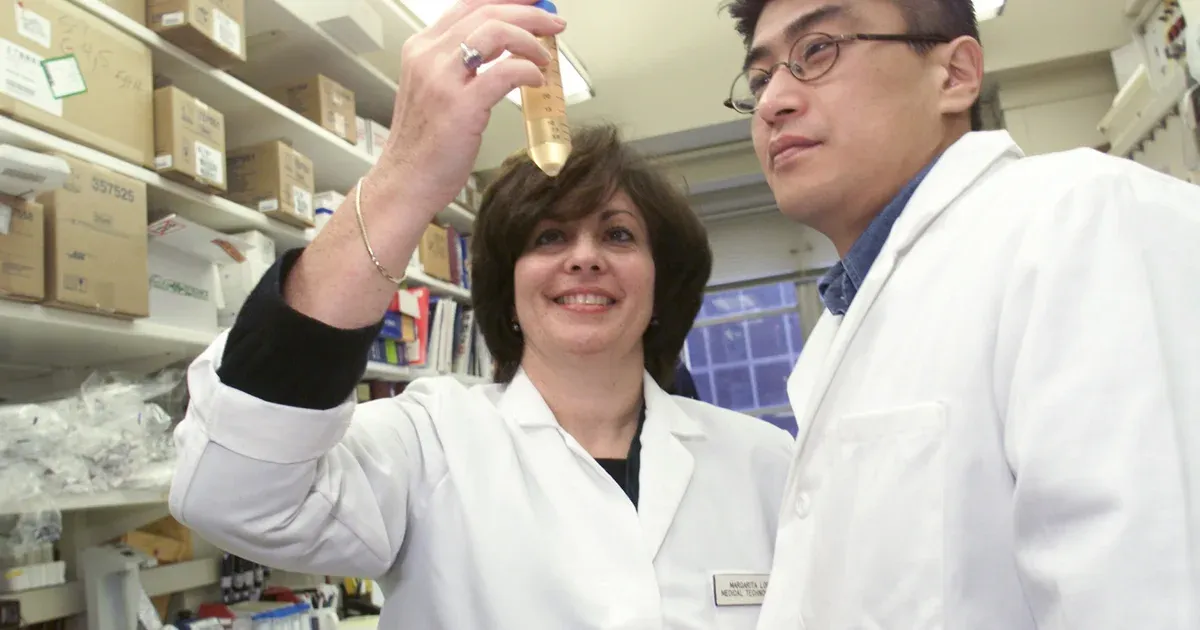
A UCLA-led team has tested a blood assay designed to detect methylation signals associated with liver, lung, ovarian and stomach cancers while using much less sequencing data than many genome-wide methods. The assay, called MethylScan, was evaluated with plasma from 1,061 people and reported in the Proceedings of the National Academy of Sciences.
The study presents a potentially less data-intensive way to analyze cell-free DNA, the small fragments of genetic material circulating in blood. It also explores whether the same assay can distinguish several chronic liver diseases. These are early validation results, not evidence that MethylScan is ready for annual population screening or able to diagnose cancer by itself.
The distinction matters. The main performance estimates came from repeated cross-validation within the study dataset rather than from a separate prospective screening trial. A positive signal would still require clinical investigation to determine whether cancer is present and where it is located.
How MethylScan Reduces the Sequencing Burden
Cancer can alter the chemical tags attached to DNA. MethylScan searches for those methylation changes in cell-free DNA. The researchers designed a capture panel covering 154,028 genomic regions and used methylation-sensitive restriction enzymes to remove much of the low-methylation background before sequencing.
This enrichment step concentrates the signal in selected regions instead of attempting to read the entire circulating methylome at extreme depth. The study used an average of about 5.3 gigabases of sequencing data per plasma sample, which the authors described as an effective depth of roughly 300 times across the targeted regions.
The paper estimates that the sequencing component could cost less than $20 per sample if data can be generated for less than $4 per gigabase. That figure is not the price of a finished clinical test. Blood collection, laboratory preparation, capture reagents, quality control, computation, professional interpretation and follow-up care would all add cost.
For marker discovery, the team also analyzed 189 matched pairs of tumor and nearby normal tissue from the four cancer types. The resulting cancer model used 15,155 methylation regions, reduced computationally to 500 features before classification. An analytical dilution experiment detected tumor-derived signal at a 0.05% spike-in level, but that laboratory limit of detection should not be confused with clinical sensitivity in patients.
What the Cancer Results Showed
The multicancer analysis included 460 samples from people with cancer and 401 noncancer samples from general hospital visitors. Researchers ran stratified fivefold cross-validation 50 times. Across all stages, the model reached an area under the receiver operating characteristic curve of 0.938. At a fixed specificity of 98%, sensitivity was 63.3%.
For early-stage cancers, the reported area under the curve was 0.916 and sensitivity was 55.3% at the same 98% specificity. Stage I sensitivity was 54.4%. High specificity limits false-positive results in the tested comparison group, but it does not reveal how the assay would perform when cancer is rare in a general screening population.
The model also attempted to predict the tissue of origin among the four cancer types. It assigned the correct cancer type in 91.7% of cancer samples that first exceeded the detection threshold, and in 89.8% of detected early-stage cases. Those percentages exclude cancers the initial detector missed and should not be read as accuracy across every tested participant.
Cross-validation is a legitimate way to estimate performance while developing a model, and the researchers repeatedly separated training and test folds to reduce instability. It is still weaker evidence than locking the model and testing it in a new population recruited for the intended clinical use. Differences in age, ancestry, other illnesses, sample handling and cancer prevalence can change real-world results.
Liver Surveillance Produced a Separate Result
The investigators separately studied 91 people with liver cancer and 157 high-risk participants without liver cancer. In this cohort, a liver-specific model produced 79.6% sensitivity at 90.4% specificity. Sensitivity for early-stage liver cancer was 76.3%.
This comparison is relevant because people with cirrhosis and some chronic liver diseases already undergo surveillance for hepatocellular carcinoma. It does not show that a blood assay can replace ultrasound, alpha-fetoprotein testing or diagnostic imaging. A future trial would need to compare the entire screening pathway, including cancers found, cancers missed, false alarms and the consequences of follow-up procedures.
MethylScan was also used to classify alcohol-associated liver disease, metabolic dysfunction-associated steatotic liver disease, viral hepatitis and autoimmune liver disease. Only 62.4% of the 157 high-risk participants met the model's confidence rule. Among that selected group, classification accuracy was 84.7%. The smaller alcohol-associated and metabolic-disease groups performed less well, so the result does not support broad organ-disease diagnosis from a single blood draw.
The study also found methylation patterns associated with self-reported race, an observation the authors identify as a possible confounder for disease prediction. That makes balanced recruitment and external validation especially important. A model can appear accurate if it learns demographic differences that happen to correlate with disease in its development dataset.
A Screening Test Needs More Than a Strong Curve
The next decisive evidence would come from a prospective study in the population where the test is meant to be used. Researchers would need to predefine thresholds, process samples consistently and report performance by cancer type, stage and demographic group. They would also need to track what happens after a positive result.
Positive predictive value will be central. Even a highly specific test can generate more false positives than true positives when used for uncommon cancers in people without symptoms. Follow-up imaging and biopsies can carry financial, physical and psychological costs, so a useful screening program must show that benefits outweigh those harms.
The present study supports further development because it combines targeted methylation enrichment with relatively modest sequencing volume and reports measurable signals in four cancers. It does not yet show reduced cancer mortality, clinical benefit or safe population-wide implementation. The credible conclusion is narrower: MethylScan has produced promising retrospective performance that now requires independent, prospective testing before it can be judged as a screening tool.